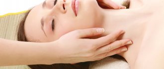
How long should the neck be
Of course, there is no need to talk about any calculations in centimeters. The length of the neck - like all other parts of the body - depends on the height and general proportions of the figure.
The German anthropologist Julius Kohlmann concluded at the beginning of the last century that if we take the entire height of a proportionally built person as 100 units, then the height of the head should be 13 parts, the height of the head with the neck 20 parts. That is, the neck makes up 7% of our height.
But, of course, these indicators are the “average temperature in the hospital”: a lot depends on the person’s build type. In people of the thin-boned asthenic type, the neck is thinner and longer than in normosthenics, and in people of the broad-boned type, on the contrary, it is shorter.
Neck length is the distance from the angle of the lower jaw to the middle of the collarbone.
There is a special test that can be used to measure the length of the neck and determine whether a man's neck is too short or long. The ideal proportion is when the hollow (where the chin meets the neck) is at a distance of four fingers from the protruding bones of the collarbones. If the distance is greater, the neck is considered long, if less, the neck is considered short.
In a word, it is impossible to calculate the ideal neck length in centimeters; you shouldn’t even try. It is much more correct to apply the harmonizing approach of Andrei Iskornev, according to which the beauty of a person is not in the beauty of individual parts of his body, but in how harmoniously they relate to each other.
Anatomy of the neck: vertebrae, muscles, vessels
The human neck is the part of the body that connects the head and body. Its upper border begins at the edge of the lower jaw. In the trunk, the neck passes through the jugular notch of the manubrium of the sternum and passes through the upper surface of the clavicle. Despite its relatively small size, there are many important structures and organs that are separated by connective tissue.
blood flow
The vessels of the neck provide the outflow of venous blood from the head and neck. They are represented by the external and internal jugular vein.
Blood enters the external vessel from the back of the head in the ear area, the skin above the shoulder blade and the front of the neck. A little earlier than the clavicle, it connects with the subclavian and internal jugular vein.
The latter eventually develops into the former at the base of the neck and divides into two brachiocephalic veins: the right and left.
The vessels of the neck, and especially the internal jugular vein, play an important role in the processes of hematopoiesis. It originates at the base of the skull and serves to drain blood from all vessels of the brain.
Its tributaries in the neck area are also: superior thyroid, lingual facial, superficial temporal, occipital vein.
The carotid artery passes through the neck area, which has no branches in this area.
The lymph nodes
The anatomy of the neck also includes part of the body's lymphatic system. In this area it consists of deep and superficial nodes. The anterior ones are located near the jugular vein on the superficial fascia.
The deep lymph nodes of the front part of the neck are located near the organs from which lymph flows out, and have the same names with them (thyroid, preglottic, etc.). The lateral group of nodes consists of the retropharyngeal, jugular and supraclavicular nodes, next to which the internal jugular vein is located.
The deep lymph nodes of the neck drain lymph from the mouth, middle ear and pharynx, as well as the nasal cavity. In this case, the fluid first passes through the occipital nodes.
The structure of the neck is complex and thought out by nature down to every millimeter. A set of plexuses of nerves and blood vessels connects the work of the brain and the periphery. In one small part of the human body all possible elements of systems and organs are located: nerves, muscles, blood vessels, lymphatic ducts and nodes, glands, spinal cord, the most “mobile” part of the spine.
Source: https://FB.ru/article/248329/anatomiya-shei-pozvonki-myishtsyi-sosudyi
Causes of sprains and dislocations
- The main reason: injury received during a sudden movement or an unsuccessful fall. This type of injury occurs in accidents or falls from a height. The risk group for getting it includes children and people with weak development of the muscular corset.
- Overexertion during training and physical labor, when the body cannot withstand the load, also leads to dislocations and sprains.
- Sets of exercises performed without warming up and stretching also lead to damage to the ligaments of the neck and its muscles.
- Pain often begins after an uncomfortable sleeping position.
- The disease can be caused by drafts and general hypothermia of the body, which provokes myositis.
For any pain symptoms in the cervical spine, the patient needs to consult a doctor and undergo a medical examination to rule out serious neck injuries (for example, a fracture).
Unfortunately, it is difficult to protect yourself from injuries, but by following a number of preventive measures, a person will be able to reduce the incidence of sprains of the neck and its ligaments.
- Shortly before physical activity, you should do a mini-warm-up for the neck muscles. It will make them more elastic and improve blood circulation.
- Protect yourself from drafts indoors, and in winter, dress warmly to prevent your neck from getting hypothermic.
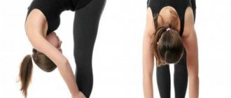
- The best way to strengthen the muscle corset in the cervical spine is to regularly perform a set of exercises to stretch the cervical spine. They will make the muscles stronger and also serve as a preventive measure for many diseases of the cervical spine, including hernia and osteochondrosis.
The complex is performed slowly, without jerking. The number of approaches increases gradually. Over time, the neck will become more flexible and stronger, but this will not happen immediately. Starting position: the patient sits on a chair. You should straighten your neck while inhaling, fix it for 5–7 seconds in the main position of the exercise, and bend it while exhaling.
- The patient sits on a chair, relaxes the whole body, lowers his arms along the body. The head slowly leans back. Fixes for 5 seconds and returns to its original position. Done 9–10 times
- Repeat the starting position. Head tilts will be performed downwards. The person presses his chin to his chest as much as possible and returns to his original position. The repetition is done 10 times.
- A person sitting on a chair turns his head to the maximum amplitude, first to the left, then to the right. 10 turns are made in each direction.
- The patient relaxes the body, sits on a chair, lowers his arms along the body. The head is thrown back. You need to stretch your left ear to your left shoulder, touching it. The same is done with the right ear. Repeat: 10 times.
- From the starting position, we do the following exercise, this time we place our hands at head level. We strain the muscles of the cervical region, bend over and press our left temple into our left palm for 5 seconds. Then we do similar bends with pressing with the right temple. Number of approaches: 3 on each side.
- The chin is pressed to the chest. The head carefully turns to the left, then to the right. The chin does not come off and should slide over the chest. The exercise is done at least 5 times.
- The patient writes out numbers from 1 to 9, moving only his nose. Then he writes the numbers from 1 to 9 in reverse order. With the help of such manipulations, the vestibular apparatus is also perfectly trained.
- Both palms are placed on the forehead, so that one lies on top of the other. A person presses his forehead onto his folded palms, while simultaneously straining his neck muscles. Do 3 sets of 5-7 seconds. Then the palms move to the back of the head, and the same manipulations with the back of the head are repeated.
- Stretching of the cervical vertebrae. This exercise is done very carefully; you may feel dizzy. The fingers are interlocked in a “lock” and placed on the back of the head, while the palms cover the neck. The chin rests on the elbows. The elbows are brought together. The elbows, chin and “lock” of clasped fingers are carefully raised and fixed in this position for 10 seconds.
- Place your palms on the back of your head and lightly press on your head for 20 seconds. Gradually, the time of load on the neck muscles increases. In the first seconds, it is recommended to apply light pressure, which increases towards the middle of the exercise and decreases again towards the end of the exercise.
- The person stands up, his back should be straight, his legs apart. The palms are placed on the chest area, below the larynx. Inhale, hold your breath for 3-5 seconds, exhale. As you exhale, apply pressure with your hands on the neck muscles. With each approach, the arms move down a little. The end point of folding the arms is the mammary glands.
- The person lies on his back, placing a cloth roll under his neck. Hands are placed on the neck so that the thumbs are under the chin, and all other fingers are collected in a “lock” at the back of the head. The head moves forward a little, bending towards the chest. The head is slowly pulled up along the trajectory of the spinal column. Execution time does not exceed 20 seconds. If desired, this exercise can be done at work. In this case, the person does not get up from the chair, but the head is gently pulled up with the help of the hands.
We'll surprise you: it often happens that men and women with short necks are not short-necked at all. After all, visually the length of the neck depends on many factors, among which its anatomical length is far from the first place.
So, why is the neck short or, more accurately, looks short?
The first reason is posture.
Surely you have often noticed the swan necks of beauties from portraits of the 18th and 19th centuries. Have you forgotten to look at their straight backs, proud head carriage and sloping shoulders? Dropped shoulders will immediately visually add several centimeters of length to your neck. But raised shoulders and a stoop “eat up” the same few centimeters.
Correct posture makes your neck longer
A very illustrative example of how the position of the shoulders and head - that is, posture - changes the perception of neck length is given to us by the famous women of the Padaung people, who have been wearing brass rings around their necks since childhood. An adult woman can have up to two dozen such rings and her neck becomes incredibly long.
The rings deform the upper thoracic region, lowering the shoulders and collarbones, and on the other hand, teach their owner to constantly live with her chin raised high. The result is the longest female neck on planet Earth.
A Padaung woman's neck is no longer than ours
Of course, no one is suggesting that you use the Padaung method. We suggest you just remember to keep your shoulders down and your chin up. The miniature device Lumo Lift will help with this. It attaches to your lapel and vibrates when you slouch.
The second reason why your neck appears shorter than it is is excess weight.
Excessive volume of the face - round cheeks, double chin, jowls, fat deposits on the neck itself - this is how a short, thick neck is visually formed, and when combined with raised shoulders (a frequent companion to a computer and sedentary lifestyle), it simply becomes invisible. In this case, it is enough to reduce weight to make the neck thinner and longer.
The result “before” and 10 days “after” the operation. Surgeon: Iskornev Andrey.
The third reason is age-related changes.
Loose skin under the chin, the formation of a goiter and the so-called turkey neck make the angle between the neck and chin less pronounced, and the outlines of the neck become blurred. As a result, it appears much shorter than it actually is.
Photos “before” and 12 hours “after” the operation. Surgeon: Vasiliev Maxim.
The fourth reason is congenital anatomical features.
Removal of Bisha's lumps, chin endoprosthetics with a Porex implant (USA), laser liposuction of the chin, thread lifting of the lower third of the face. Surgeon - Iskornev A.A.
The structure of the human neck from the front and back: lymph nodes and muscles of the neck, their diseases and treatment
One of the most important parts of the human body is the neck. It connects the head and torso.
Regions and boundaries
The upper border of the neck coincides with the lower edges of the jaw and bony auditory canal, as well as with the upper border of the occiput. The lower border runs along the jugular fossa on the front surface of the neck, along the upper side of the clavicle and along the upper line of the processes of the shoulder blades.
Form
The shape of the neck is individual for each person, it depends on age, gender, weight and muscle corset. In all people, without exception, the neck has a cylindrical shape, the skull serves as the border on top, and the shoulder girdle below.
Organs
Inside the neck are many vital organs and anatomical structures.
Organs located inside the neck:
- Larynx. Performs a protective and protective function. Protects the respiratory tract from the penetration of foreign substances and bodies.
- Pharynx. Takes part in the processes of speech formation and breathing, and also plays a role in the conduction of food. In addition, it has a protective function.
- Trachea. An important respiratory organ that conducts atmospheric air to the lung sacs. It also helps in sound production by conducting air to the vocal cords.
- Connective type fabric. It is necessary to perform protective and support functions.
- Thyroid. One of the main glands that produce hormonal substances necessary for normal metabolism.
- Esophagus. This organ of the digestive system pushes the bolus of food into the stomach for further processing.
- Spinal cord. Its functions are to generate autonomic and motor reflexes; in addition, it is a kind of “bridge” connecting the brain with the peripheral part of the nervous system.
- Subcutaneous fatty tissue. Performs the function of protection and shock absorption, while promoting thermal insulation and energy supply to the internal organs of the neck.
Here you will find a similar article on the topic “What does an ultrasound of the neck show and how to prepare for it?”
Neck parts
In the human neck, it is conventional to distinguish four sectors or areas:
- Back of the neck.
- Lateral or lateral region of the neck.
- Sternocleidomastoid region of the neck.
- Anterior neck area.
Each of the above areas has its own specific structure in accordance with the functions it performs. And in each area there are muscles, organs, and networks of the circulatory and nervous systems that limit it.
Neck bones
The mobility of the neck is due to the spinal column passing through it. The human spine consists of 33-34 vertebrae, but the cervical spine includes only 7. A distinctive feature of the cervical vertebrae is its small and short bodies.
This is explained by the fact that, compared to other sections, the cervical vertebrae bear the least amount of load. But, despite this, it is the neck region that is more susceptible to various injuries and sprains, since the muscle corset is quite weak.
The cervical vertebrae have transverse processes through the openings of which pass the vessels of the circulatory system. These vessels bring oxygenated blood to the brain.
Atlas vertebra
The first cervical vertebra of a person is called “Atlas”. This is due to the fact that it performs a rather important function of connecting the skull with the spinal column.
Unlike all other vertebrae, the atlas does not have a body. In this regard, the opening in the vertebra is noticeably enlarged, and both arches (posterior and anterior) are connected to each other using lateral masses.
On the anterior side of the anterior arch there is a tubercle, and on the posterior side there is a tooth fossa, with the help of which the atlas is connected to the second cervical vertebra.
The atlas does not have a spinous process; there is only a posterior tubercle on the posterior arch, which is an underdeveloped process.
On the lateral surfaces of the atlas there are articular surfaces, both on the upper and lower sides. The upper articular surfaces form the atlanto-occipital joint by connecting with the condyles of the occipital bone.
The lower ones connect to the upper articular surfaces of the second cervical vertebra and form the lateral atlantoaxial joint.
Axis
Axis or epistropheus is the second cervical vertebra of a person. Its distinctive structural feature is the presence of a process (tooth) that extends upward from the vertebra. This process has an apex and two articular surfaces.
The anterior surface connects with the tooth fossa of the posterior surface of the atlas and forms the median atlanto-axial joint. The posterior surface of the axis attaches to the transverse ligament of the first cervical vertebra.
The upper articular surfaces of the axis are located on the lateral sides of its body. The superior surfaces articulate with the inferior surfaces of the first cervical vertebra and form the lateral atlantoaxial joints.
The inferior surfaces of the axis are necessary for the connection of this vertebra with the third cervical vertebra.
We wrote about what is located in the left side under the ribs in front of a person here.
Muscles of the cervical region
The function of the muscular corset of the neck is to maintain the position of the head in space, as well as to carry out motor movements of the neck and head. In addition, muscles are involved in swallowing and generating sounds.
All muscles are divided into two groups: native and alien muscles. Their main difference is that the alien muscles are associated with the movement of the upper limbs, and their own muscles are necessary for the movement of the head and torso.
Own muscles:
- Longus colli muscle. Its main function is aimed at flexing the neck and body, the principle of operation is the opposite of the principle of the spinal muscles.
- Longus capitis muscle. The function performed is exactly the same as that of the longus colli muscle.
- Anterior, middle and posterior scalene muscles. These muscles are involved in the breathing process, namely, during the inhalation stage, the ribs rise with the help of these muscle fibers.
- Sternohyoid, omohyoid, sternothyroid, thyrohyoid and geniohyoid muscles. These muscle fibers pull the larynx and hyoid bone down.
Alien Muscles:
- Mylohyoid, stylohyoid and digastric muscles. They, due to the fact that they pull the larynx and hyoid bone up, are able to lower the lower jaw down.
- Subcutaneous muscle of the neck. Its main function is to protect the saphenous veins from excessive pressure. This occurs due to the stretching of the skin of the neck by this muscle.
- Sternocleidomastoid muscle. There are two types of contraction of this muscle: unilateral and bilateral. In the first option, the head tilts to the side and the face turns upward. With bilateral contraction, the head tilts back and rises upward. This contraction is also necessary to support the head in an upright position and to inhale.
Fascia of the neck
Fascia is a connective tissue sheath that covers muscles, tendons, organs, and bundles of nerves and blood vessels.
According to the classification of Doctor of Medicine, Academician, Professor V.N. Shevkunenko. There are five fasciae in the neck:
- Superficial fascia.
- Proprietary fascia of the neck.
- Scapuloclavicular fascia.
- Intracervical fascia.
- Prevertebral plate.
Blood flow in the neck
The circulatory system in the neck area is formed by an interweaving of vessels of various types, the main task of which is to ensure the delivery of blood to and from the brain. There are arterial and venous systems.
The arterial circulatory system includes:
- Common carotid artery. It, in turn, is divided into internal, which carries blood to the orbital area of the head, and external, which promotes blood flow to the facial and cervical region.
- Subclavian artery.
The venous system includes the following vessels:
- Thyroid vein.
- Anterior jugular vein.
- Internal jugular vein.
- External jugular vein.
- Subclavian vein.
The function of veins, unlike arteries, is to ensure the outflow of blood from the brain.
Nerve plexus of the neck
The cervical nerve plexus is formed by the four upper cervical spinal nerves, namely their anterior branches, which are connected by three arcuate loops.
The nerve plexus of the cervical region is located on the anterior and lateral surface of the deep cervical muscles, and the sternocleidomastoid muscle closes it on top.
The nerve plexus of the neck includes the following nerve branches:
- Muscle nerves. Their main function is to establish a relationship between the surrounding muscles and the central nervous system. These nerves are of the motor type.
- Cutaneous nerves. They belong to the sensitive type of nerves.
- Phrenic nerve. The motor fibers of this nerve innervate the diaphragm directly, and the sensory fibers innervate the peritoneum.
Lymph nodes
The lymphatic system as a whole performs a protective function. Lymph nodes are peripheral organs that filter all lymph passing through the body.
There are several groups of lymph nodes in the cervical region, including:
- chin;
- parotid;
- supraclavicular;
- subclavian;
- retropharyngeal, etc.
Depending on the location of the lymph nodes, they can perform a protective function for certain organs and tissues.
In healthy people, the lymph nodes are not visible or palpable. Only with various types of diseases do the nodes of the lymphatic system become noticeably larger and are easy to detect with the naked eye.
Diseases in the neck area
Often the cervical region is vulnerable and susceptible to a variety of inflammatory diseases. Very often, boils and carbuncles appear on the back of the neck.
Most often, such formations occur in places of strong friction with clothing, or in especially sweaty areas where there is little hair.
Inflammation of the lymph nodes in the neck is also common. There is a chronic form of lymphadenitis, in which no purulent processes occur, as well as an acute form of the disease.
It is in the acute form that immediate surgical intervention is often necessary, since the lesion grows very quickly, followed by massive suppuration and tissue necrosis.
Among tumor diseases, benign formations such as angiomas, fibromas, lipomas, neurofibromas, etc. are distinguished. Malignant tumors include cancer of the lip, thyroid gland, lymphosarcoma, etc. Treatment options include surgery and radiation therapy.
In addition to all of the above, diseases of the spine can develop, for example, osteochondrosis, and inflammation of the neck muscles is also common.
Competent treatment of diseases in the neck area
Very often you can hear complaints about pain in the neck; in most cases, such sensations are associated with inflammation of the neck muscles.
Below are several folk remedies that will help reduce pain and relax the neck muscles:
- Various warming ointments have a strong effect. You need to mix about 2 tablespoons of butter and a teaspoon of horsetail in powder form. This mixture is applied to the inflamed area, cotton wool is applied, wrapped in cling film and wrapped in warm cloth. Leave it like this overnight, and the next day the pain should disappear.
- You can also use cabbage compresses. To do this, rub the cabbage leaf with laundry soap and sprinkle with baking soda. This side up should be applied to the neck and wrapped with a warm scarf. The next day the neck muscles will be relaxed.
In addition, exercises to strengthen the neck muscles and massage this area will be good prevention.
Below are useful exercises as an example:
- Stand up straight, hands at your sides, carefully tilt your head from side to side without sudden movements.
- The starting position is the same, turn your head from side to side.
- Sitting on a chair, place your palms on your forehead, gently pressing with your hands, try to overcome the force and pressure, hold for a few seconds, then relax.
- In the same way, while sitting on a chair, press your palm on your temple and try to overcome the pressure, hold for a few seconds and relax. Repeat on the other side.
Source: https://spinaspina.com/diagnostika/stroenie-shei.html
What to do if you find a lump on your neck
If you have a lump on the front of your neck, you need to clearly assess the problem and not give in to panic. If it formed in a short time, then quite soon the process may reverse. It is important to keep the pine cone clean and not touch or damage it.
But when you begin to notice symptoms such as thickening or an increase in the diameter of the lump in the absence of other infections, the duration of remission, the accompanying deterioration in well-being or discomfort in the area of the growth, it is necessary to urgently consult a specialist. You should contact a therapist, dermatologist, surgeon, oncologist, infectious disease specialist or endocrinologist.
It is these specialists who will advise you on how to remove a lump on your neck, depending on the cause of its occurrence.
Neck areas
The upper border of the neck is drawn from the chin along the base and posterior edge of the ramus of the mandible to the temporomandibular joint, and continues down and posteriorly (through the apex of the mastoid process of the temporal bone) along the superior nuchal line to the external protrusion of the occipital bone.
The lower border of the neck runs from the jugular notch of the sternum along the upper edge of the clavicle to the top of the acromion and further to the spinous process of the 7th cervical vertebra. The following areas of the neck are distinguished: anterior, sternocleidomastoid - right and left, lateral - right and left and posterior.
The anterior region of the neck (anterior triangle of the neck) has the shape of a triangle, the base of which faces upward. This area is bounded above by the base of the lower jaw, below by the jugular notch of the sternum, and on the sides by the anterior edges of the right and left sternocleidomastoid muscles. The anterior midline divides this area of the neck into the right and left medial neck triangles.
The sternocleidomastoid region is paired, corresponds to the location of the muscle of the same name and extends in the form of a strip from the mastoid process above and behind to the sternal end of the clavicle below and in front.
The lateral region of the neck (posterior triangle of the neck) is steamy and has the shape of a triangle, the apex of which faces upward; the region is located between the posterior edge of the sternocleidomastoid muscle anteriorly and the lateral edge of the trapezius muscle posteriorly. Bounded below by the collarbone.
The posterior region of the neck (nuchal region) on the sides (right and left) is delimited by the lateral edges of the corresponding trapezius muscles, the superior-superior nuchal line, the inferior-transverse line connecting the right and left acromions and drawn through the spinous process of the VII cervical vertebra. The posterior midline divides this area of the neck into right and left parts.
Within the anterior and lateral regions of the neck, a number of triangles are distinguished, knowledge of which is of great practical importance, especially during surgical interventions. In the anterior region of the neck, on each side, three triangles are distinguished: carotid, muscular (scapular-tracheal) and submandibular.
The carotid triangle is bounded posteriorly by the anterior edge of the sternocleidomastoid muscle, anteriorly and inferiorly by the superior belly of the omohyoid muscle, and superiorly by the posterior belly of the digastric muscle.
The muscular (scapulotracheal) triangle is located between the anterior edge of the sternocleidomastoid muscle posteriorly and inferiorly, the superior belly of the omohyoid muscle superiorly and laterally, and the anterior midline medially.
The submandibular triangle is bounded below by the anterior and posterior bellies of the digastric muscle, and above by the body of the mandible. In the area of this triangle lies the salivary gland of the same name.
Within the submandibular triangle, a small but very important for surgery lingual triangle, or Pirogov’s triangle, is distinguished. It is bounded anteriorly by the posterior edge of the mylohyoid muscle, posteriorly and inferiorly by the posterior belly of the digastric muscle, and superiorly by the hypoglossal nerve.
The entire area of the triangle is occupied by the hyoglossus muscle, by spreading the fibers of which, you can find the lingual artery.
In the lateral region of the neck, the scapuloclavicular triangle and the greater supraclavicular fossa are distinguished.
The scapuloclavicular triangle is small in size. Located directly above the middle third of the clavicle, bounded below by the clavicle, superiorly by the inferior belly of the omohyoid muscle, and anteriorly by the posterior edge of the sternocleidomastoid muscle.
The lesser supraclavicular fossa is a well-defined depression above the sternal end of the clavicle, which corresponds to the space between the lateral and medial legs of the sternocleidomastoid muscle.
In the neck area, the sternocleidomastoid region is also distinguished. It corresponds to a prominently expressed elevation formed by the muscle of the same name. The interscalene space extends between the middle and anterior scalene muscles; from below it is limited by the first rib.
The subclavian artery and trunks of the brachial plexus pass through its space. The prescalene space is limited anteriorly by the edges of the sternothyroid and sternohyoid muscles, and posteriorly by the anterior scalene muscle. The subclavian vein passes through this space.
The structure of the human larynx
The larynx is attached by muscles to the hyoid bone and connects the nasopharynx with the lower respiratory tract - the trachea and lungs. The shape of this organ is provided by a system of cartilages that form a flexible, movable tube.
The cricoid cartilage underlies the larynx, the thyroid cartilage serves as a frame, and the epiglottic cartilage acts as a lid, protecting the airways from chewed food during swallowing.
Paired cartilages (cuneiform, arytenoid, corniculate) strengthen the larynx, help it narrow and expand.
Look at the photo of how the human throat works:
Inside, the larynx is similar to an hourglass, in the middle of which there are elastic ligaments that form an opening for the passage of air - the slit.
The tone of the voice and its individual color are regulated by the length of the chords according to the principle: the shorter the length, the higher the timbre. The larynx is in constant motion: when exhaling and swallowing or singing, it rises, and when inhaling, producing low sounds, it lowers.
The larynx and pharynx are associated with the breathing process: inhaled air from the nose passes through these sections and rushes further into the trachea, to the lungs. Together they participate in the reflexive process of swallowing. The tissues of the pharynx protect against infection, and the structure of the larynx protects the airways from food getting into them. The larynx “gives birth” to the voice, and the pharynx strengthens it.
Here you can see a diagram of the structure of the human throat:
If you have questions for your doctor, please ask them on the consultation page. To do this, click on the button:
Ask a Question
Source: https://neuro-orto.ru/lechenie/oblasti-shei.html
Photos "before" and "after"
Neck lift, results before and 12 hours after! After the operation: the oval and contour of the lower jaw are emphasized, the jowls are corrected, the double chin disappears, the chin does not sag when the head is tilted! Performed by surgeon: Maxim Vasiliev.
Performed facelift-smas, lipofilling, face, co2-lifting. Before and 9 days after. Performed by surgeon: Maxim Vasiliev.
Hollywood neck plastic surgery. Photos were taken “before” and 7 days “after”. Surgeon: Iskornev Andrey.
Neck lift. Author's technique. “Before” and 7 days “after” the operation. Performed by surgeon: Maxim Vasiliev.
Neck lift using dynamic platysmoplasty - the chin will not sag even when tilted. Result 10 hours after surgery. There is swelling and markings. Surgeon: Vasiliev Maxim.
Platysmaplasty with installation of a Medpor chin implant. Photos were taken “before” and 12 days “after”. Surgeon: Vasiliev Maxim.
Neck lift, results “before” and 12 hours “after”. Author's technique. Performed by surgeon: Maxim Vasiliev.
Platysmoplasty. Surgeon: Iskornev Andrey.
Neck lift (author's technique). Photos “before” and 2 days “after”. Performed by surgeon: Vasiliev M.N.
The operation was performed by Iskornev A.A.
Platysmoplasty. Surgeon - Andrey Iskornev.
Symptoms and diagnosis
If a person has “strained” his neck, then he begins to experience acute pain in the damaged area, which intensifies with head movements (turns, bends). Muscle tone is increased, and upon palpation of the painful area, discomfort intensifies. The damaged (stretched) area turns red and swells. Neck movement is limited, and the patient is forced to look for a more comfortable position for the head, which temporarily relieves discomfort.
In young children, it is more difficult to detect damage. Parents should pay attention if the child is lethargic, the neck area is swollen, its movement is limited, and the head is turned unnaturally. In difficult cases, due to injury, for example, an accident or fall, the baby may lose consciousness. A child older than one and a half years will already be able to independently complain to his parents about pain.
Causes of shortened neck and sagging face
Neck protraction is the movement of the neck forward. With age, a healthy spine begins to “sag”, the vertebrae become deformed, and as a result, hyperlordosis of the neck is formed - a deflection of the neck inward. The vertebrae collapse into the neck, and its length decreases.
You can ask to have your photo taken in profile; if there is a shortening of the neck, then your back will be round, your shoulders will be concave into your chest, and your withers (hump) will most often be visible.
In addition to shortening the neck in length, its visual shortening can also be caused by spasm of the shoulder muscles. “Gadget neck” is formed from incorrect posture, from often bowing the head down. Shoulder muscle spasms can also be caused by tension, nervous stress, etc.
With cervical hyperlordosis, when the neck arches too much at the back and, accordingly, protrudes in front, it is impossible to maintain a clear oval of the face and a taut chin.
Train your neck muscles! They are the “foundation” of the face! If the skin on your neck hangs down the front, always look for the problem at the back of your neck.
If the neck is in a state of “protraction” - it moves forward, then it needs to be moved to the opposite state - retraction, lengthening the suboccipital muscles, “pulling out” the vertebrae that have sunk inside.
It is useless to do exercises for double chin, “turkey skin” and wrinkles on the side of the neck; you need to eliminate the cause of sagging, i.e. eliminate hyperlordosis and neck protraction.
Treatment
First aid for sprains:
- Complete rest of the damaged area. The patient is placed on a flat surface and a cloth cushion is placed under the neck. In this position, the load on the spine is more even, and the pain subsides.
- To relieve swelling and pain, apply a cold compress to the neck. Ice and snow can only be used by wrapping it in a piece of cloth or gauze.
- The use of anti-inflammatory medications and the use of pain-relieving ointments.
- If the damage is the result of an accident (fall, accident), call a doctor immediately.
At home
The doctor diagnoses the damage and prescribes a course of treatment. Typically, treatment for sprains is done at home. On average, the recovery period is from 2 to 3 weeks. For treatment at home, compresses are used. The patient is recommended to take painkillers and undergo a course of physical therapy. Neck massage in combination with physical therapy will help restore the neck.
Exercise “Neck Protraction – Neck Retraction”
Neck protraction is a forward movement of the neck, which is accompanied by increased cervical lordosis and, accordingly, a change in pressure on the surface of the vertebra. Neck retraction is the exact opposite movement. In this case, the forehead and chin are in the same projection.
Make a neck retraction movement: move the lower jaw towards the spine (left). We make a movement for 10 counts: forward - backward in dynamics. And back: a delay of 10 counts.
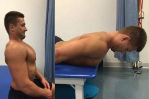
Neck retraction can be complicated by lying on the stomach, chest extended beyond the edge of the bed, arms along the body, shoulder blades in the middle position between full reduction and removal from the spine (right). Care should be taken to avoid neck flexion during retraction.
See 3 exercises for correcting protraction to the retraction position from Inna Nefedovskaya
The video shows 3 exercises, but you can order the whole set of exercises from the online course “Posture and neck, like in youth”, click on the button