Breast diseases are the most common pathology faced by middle-aged and older women. Almost 15% of them are cancer. This means that effective preventive screening of the population is necessary to detect the disease at an early stage of development. The main one is mammography.

Breast diseases are the most common pathology faced by middle-aged and older women.
Breast mammography is an X-ray examination method that has a high sensitivity and specificity. This examination is available to all segments of the female population, is organized en masse in medical institutions and is the “gold standard” for detecting tumors.
Indications
Mammography is performed on all older women at regular intervals.

A palpable formation in one of the glands is an indication for screening.
Also, an extraordinary study is prescribed if there are certain indications, such as:
- palpable hard or soft formation in one of the glands;
- visual deformation of the nipple or any other area;
- the appearance of blood when pressing on the nipple;
- local pain, signs of swelling, change in skin color, violation of symmetry on both sides.
Women of the young age category are referred for mammography under the following conditions:
- risk group for breast cancer (close relatives have been diagnosed with a malignant neoplasm);
- cancer of the uterus or ovaries;
- inability to get pregnant;
- diseases of the endocrine system;
- surgical interventions on mammary organs in the past.
Advantages
Breast ultrasound is considered an “easier” examination method compared to X-ray mammography. Ultrasound allows you to observe the dynamics of already identified tumors, changes in their size, shape and structure.
The examination allows you to assess the density and structure of the breast, as well as monitor the condition of silicone implants, if any. The absence of radiation exposure can also be considered an advantage of ultrasound examination, because this makes it possible to study the mammary gland in pregnant and lactating women.
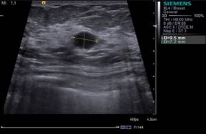
Ultrasound of the mammary glands - image
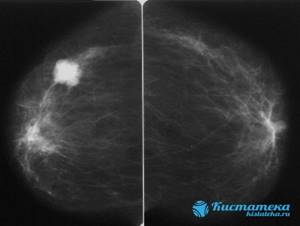
X-ray mammography is considered more accurate in examining the ducts of the mammary glands. According to the results - the images - all the structures of the breast are clearly visible, even small neoplasms are revealed. X-ray mammography is considered the gold standard for diagnosing breast cancer in all developed countries.
Did you know that ultrasound of the mammary glands must be taken on certain days of the cycle? Read more about this study and the rules for conducting it on our website.
In what cases a breast biopsy is required, you will find out here.
If malignant neoplasms of the mammary glands are suspected, a puncture of the mammary gland may be prescribed. You will find everything about this procedure at the link.
What does breast mammography show?
Mammographic imaging of the mammary glands may show cysts, mass formations (tumors), and calcifications. The general type of structure of the organ is also assessed (glandular, fibro-fatty, mixed with a predominance of one of the components). Attention is paid to the milk ducts, their structure and diameter.
Visualization of the mammary organs can show cysts, space-occupying formations (tumors), and calcifications.
Some mammographs are equipped with equipment for a needle biopsy, which is performed immediately to confirm or exclude the malignant nature of the process.
When to do a mammogram of the mammary glands?
Mammography is carried out as a preventative measure for all women over 40 years of age once every 2 years. At the discretion of local institutions for the female population over 50 years of age - once a year.
In addition, there are unscheduled indications for this type of examination.

Carrying out a mammogram while breastfeeding is completely acceptable.
During pregnancy at any stage, it is preferable to use the ultrasound method due to the absence of side effects, primarily radiation exposure to the body of the mother and fetus. Carrying out a mammogram during breastfeeding is quite acceptable, but it is not very informative for a doctor. It is recommended to use other auxiliary studies.
It is considered most correct to undergo a mammological examination on days 5-13 of the menstrual cycle, i.e. in the first phase. In the second phase, the breast tissue undergoes some hormonal changes: the ducts expand, the structure of the glandular component changes, and swelling of the soft tissues may appear. In this case, the result may be unreliable, and the examination will be endured with a feeling of discomfort and pain.
During menopause, mammary organs can be examined at any time.
Comparison of ultrasound and mammography
Thus, both methods have both positive and negative sides. To find out which is better, ultrasound or mammography, you need to compare them:
- Operating principle. Mammography is based on the use of X-rays, and ultrasound uses ultrasonic waves.
- Age of patients. Mammography is performed for women only after 40 years of age, but ultrasound can be done for everyone.
- Effect on pregnancy and infant feeding. Mammography, unlike ultrasound, cannot be performed during this period.
- Safety. During mammography, the patient receives a small dose of radiation; ultrasound examination is safe.
- Information content. It is impossible to unequivocally answer the question of what is more informative, since each of the studies can help diagnose diseases that are difficult to detect by other methods. Thus, mammography is indispensable for studying the condition of the milk ducts, and ultrasound is a means for performing a biopsy, and also allows you to assess the condition of the axillary lymph nodes.
- Soreness. When undergoing a mammogram, minor discomfort is possible, but there is none when performing an ultrasound.
- Gland size. There are difficulties in performing mammograms on women with very large or very small breasts. Unlike mammography, when performing an ultrasound, the size of the gland does not play a role.
- Accuracy of the result. With mammography, the resulting image is much clearer and the results are more accurate. Ultrasound has certain resolution limits.
- Directionality. Mammography is the main method for examining the milk ducts. Ultrasound is used to control the biopsy.
- Possibility of false result. This possibility remains when diagnosing using both methods.
To summarize the above, it can be noted that it is impossible to choose between mammography of the mammary glands or ultrasound of the mammary glands. Reviews from doctors about the diagnostic qualities of each method indicate that both of them are good in their own way. That is why these studies are complementary and, when used together, allow the doctor to make the correct diagnosis and prescribe the appropriate treatment. This is especially true for women who have reached the age of forty.
How to prepare?
No special measures are required before the manipulation. The results of mammography are not affected by food or liquid intake. The only recommendation is to avoid using cosmetics for the skin of the axillary area on the day of the study.
Before the procedure, you should remove all neck jewelry, raise your hair, and completely free your chest from clothing.
The radiologist or laboratory assistant warns the woman about all possible side effects, asks the day of the menstrual cycle, and asks about pregnancy at the time of the mammary gland examination.
To correctly interpret changes in the mammary organs, you need to have with you the protocols of previous studies (mammography, ultrasound, MRI).
Study selection
Only a doctor should determine the method of instrumental diagnosis of breast disease. After examination and palpation of the mammary glands, if the formation of compactions is suspected, additional research must be carried out.
They will provide the necessary clarifying information. At the same time, the doctor takes into account the presence of concomitant pathologies, the woman’s condition, age, phase of the menstrual cycle, gynecological history, and data on heredity.
Often doctors prescribe both research methods. They complement the information received from them, which makes it possible to make an accurate diagnosis.
If the choice is difficult to make or ultrasound or mammography did not provide a complete picture, then there is also magnetic resonance imaging, which clarifies the diagnosis completely. However, it can only be carried out in special diagnostic departments or centers, which are not yet available everywhere.
How is a breast mammogram done?
Mammography is carried out in the X-ray room, where the mammography machine is located. The woman is shown how to position herself next to him. The position depends on the type of equipment (standing or sitting). Next, a protective apron made of lead material is placed on the waist area, covering the lower abdomen. The mammary glands are placed on a plate intended for this purpose, and the chest is pressed on top with a similar plate. Next, the x-ray technician leaves the room and turns on the machine remotely. Exactly the same actions are repeated on the other side. The whole procedure takes no more than 5-10 minutes.
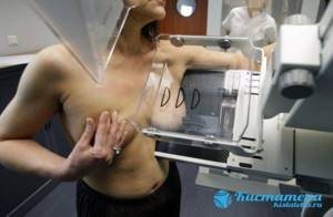
The mammary glands are placed on a plate intended for this purpose, and the chest is pressed on top with a similar plate.
If a pathology is detected, the woman may additionally undergo radiography in a different projection - lateral and/or oblique. At the discretion of the doctor, a targeted mammography is possible, when the affected area of the gland is directly imaged on an enlarged scale.
A photo with a description can be received on the same day. Some clinics are equipped with digital equipment that provides more detail in the resulting image. All information can be saved on electronic media and used at any time.
On the first day, a woman may complain that her breasts hurt after a mammogram. This is due to the mechanical pressure of the plates, does not last long and eventually goes away on its own.
Other X-ray examinations should be rescheduled on this day so as not to exceed the permissible radiation dose.
Mammography or ultrasound of the mammary glands - which is better?
Both methods, mammography and ultrasound, are highly accurate and specific for the presence of breast tumors. Ultrasound is preferable if visualization of areas located near the chest from different angles is necessary. These areas are difficult to reach for x-rays.
In addition, the ultrasound method more accurately differentiates the cystic and solid components of the tumor, identifies small pathology, and shows the presence of blood flow. It is also more informative in young females due to the increased density of organ tissue at this age. Ultrasound can be used at any stage of pregnancy.

Ultrasound is preferable if visualization of areas located near the chest from different angles is necessary.
Ultrasound allows you to analyze the size and shape of lymph nodes - an important marker for differential diagnosis between benign and malignant formations.
However, mammography makes it possible to detect single or multiple small calcifications, extensive tissue restructuring, and precise identification of the type of cyst, which is impossible with ultrasound.
Thus, both studies are complementary to each other. Combining them together significantly increases the accuracy of diagnosis and should be used when identifying pathology.
Mammography
Advantages:
- Absolute safety.
- The ability to examine the structure of the glands from all sides and in real time.
- You can examine the lymph nodes, assess the state of blood flow and neoplasms.
- It is permissible to carry out in the presence of injuries or inflammations.
- Allows you to determine the most accurate place for taking a puncture.
- The accuracy of the study is 90%. Allows you to see the slightest disturbances in the ducts at the earliest stages of their development.
- Shows places of salt accumulations (they are not visible on ultrasound).
- Provides accurate data on the location and size of the tumor.
- The accuracy of the study is 95%.
Flaws:
- Does not allow definitive diagnosis without aspiration.
- Unable to determine whether a tumor is malignant or benign.
- Requires experience, qualifications, and close attention from the doctor.
- It is forbidden to carry out frequently due to the radiation dose (small, but still).
- The lymph nodes are not clearly visible.
- Women under 40 years of age may receive distorted results due to the density of the breast tissue.
As a result, the picture is as follows: ultrasound wins in safety, mammography wins in the speed of the procedure. In terms of information content, there is no clear “winner”, since ultrasound makes it possible to evaluate the lymph nodes and ducts, and x-rays provide more accurate results about the presence of pathologies of the mammary glands. If we talk about the cost of both studies, then ultrasound will obviously be cheaper, since the price of equipment for performing it is several times lower.
Decoding the results
The results of mammography are assessed by a radiologist. The conclusion is issued on the same day. The research protocol describes the types of tissues that make up the mammary gland (fibrous, glandular, fatty), their relationship with each other, homogeneity or heterogeneity of the structure.
The shape of the organ is analyzed: round, oval, lobed or irregular. The size of the ducts is indicated (normally up to 0.4 cm in women of childbearing age, up to 0.2 in menopause), whether they are straight or convoluted, contain homogeneous or inhomogeneous contents, in which area they are better developed.
The age of the patient must be taken into account. In women over 40 years of age, involution processes begin (the glandular component is gradually replaced by fibro-fatty component). Mammography for fibrocystic mastopathy shows that during this period the glandular component predominates.

If during this period the glandular component predominates according to mammography, then fibrocystic mastopathy is diagnosed.
Dilated ducts and cysts are also clearly visible on x-rays. The doctor describes in detail their location (using a watch dial for convenience), diameter, contours, and internal contents. Types of cysts are presented as simple and complicated (festering).
The space-occupying lesion stands out quite well on the mammogram, as it is accompanied by a darkening symptom on the X-ray film. Its presence is confirmed in two projections. If visualization occurs in only one projection, then they speak of a focus of compaction in the mammary gland.
Tumors are described qualitatively by analogy with cysts, only the contours (even, uneven, presence of a capsule), density (low, mixed, high, asymmetrical), the presence and main characteristics of calcifications are analyzed in more detail.
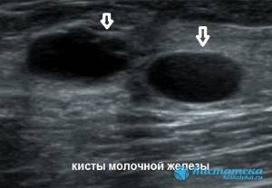
Dilated ducts and cysts are also clearly visible on x-rays.
It is now customary to assess the condition of the mammary organs according to the international system for describing and processing data BI-RADS (Breast Imaging Reporting and Data System), which is used in most countries.
The resulting changes are expressed as points. This algorithm accurately indicates to the attending physician further tactics for managing the patient and the necessary therapeutic measures. BI-RADS creates continuity between all treatment and diagnostic links that a woman goes through.
The scale includes 7 categories: 0 and 3 – cases subject to auxiliary research, re-monitoring after six months, 1 and 2 – sent to the treating gynecologist, 4 and higher – consultation with an oncologist, fine-needle biopsy is recommended.
The quality of the examination also depends on the experience and training of the specialist. In addition, there is a small percentage of false-positive diagnoses, which once again confirms the need for an integrated approach to the examination.
Signs of a benign tumor on a mammogram
Mammography can differentiate or suspect the nature of the formation. The main disadvantage is the difficulties encountered in women under 35 years of age due to the increased density of the organ parenchyma.
Among benign focal changes in the mammary gland there are lipoma, fibrolipoma, and leaf-shaped fibroadenoma.
Among benign focal changes in the mammary gland there are lipoma, fibrolipoma, leaf-shaped fibroadenoma, fibroadenolipoma, etc. They are characterized by the following signs:
- clearly defined, smooth boundaries;
- shape in the form of a circle, oval or lobes;
- homogeneous structure;
- absence of small calcifications;
- low density.
Signs of a malignant tumor on a mammogram
Breast cancer appears on mammography with certain properties:
- irregular or lobed shape;
- fuzzy (blurry), uneven contours;
- asymmetrical density;
- germination into healthy tissues;
- violation of architectonics;
- the presence of a local accumulation of small (up to 1-2 mm) calcifications in the structure or duct;
- enlargement and pathological changes in lymph nodes;
- thickening, swelling of the skin and subcutaneous fat layer above the tumor.
Other distinctive signs of malignant pathology in the mammary glands are the lack of compressibility and mobility of the formation, the vertical orientation of germination into the organ.
Thus, mammography of the mammary glands is a diagnostic technique that can establish the correct diagnosis as early, effectively, non-invasively, accurately, and easily as possible. In complex or doubtful cases, examination of the mammary organs should be carried out in combination with other methods.
Ultrasonography
The ability of ultrasonic waves to be reflected with varying degrees of intensity from structures of the body that differ in their structure formed the basis of this method of medical diagnostics. Examination of the mammary glands using ultrasound is widespread and helps establish a diagnosis in cases where it is contraindicated or impossible to examine the patient using mammography.
To carry out this method, it is enough to have an ultrasound diagnostic device, which is available in almost every clinic today. No preparation is required for the examination; patients must be in a supine position, which is much more comfortable than a mammographic examination, which is performed while standing.
A special gel is applied to the skin of the mammary gland, enhancing the adhesion and conductivity of tissues for ultrasonic vibrations. The mobility of the sensor allows you to examine the organ from all sides, in several planes. Ultrasound makes it possible to determine the intensity of blood flow in a particular area of the mammary gland, which is important for some diseases. In addition, ultrasound has no contraindications, such as pregnancy or breastfeeding, since there is no radiation, similar to an X-ray machine.
A significant drawback of examination using ultrasound not only of the mammary glands, but of any other organ, is the direct dependence of the information content of the result on the qualifications of the specialist conducting the diagnosis itself. Lack of experience and inattention to any detail can lead to an incorrect diagnosis and missing an important point. As for the examination of the mammary glands, it is possible to miss an area or overlook any piece of tissue.