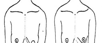
Breast diagnostics must be carried out regularly in order to prevent many diseases, among which is breast cancer. A similar procedure can be performed at home: you need to stand in front of a mirror with your arms lowered and raised (after having undressed to the waist), examine the mammary glands, turning left and right. A woman should know her own breast parameters so that she can notice minimal changes.
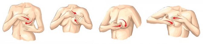
Then you need to take a horizontal position, put one hand under your head, and with the other feel your chest on the opposite side of your free limb. The palm is placed under the breast and moves upward, simultaneously feeling the mammary gland with your fingers. They also rub their fingertips around the nipples and armpits to check for any bulges. The corresponding procedure is carried out with the second breast. All movements must be performed correctly, without leaving a centimeter of the gland without attention.
Important! Such manipulations must be carried out every month during the first week after the end of menstruation.
If any deviations were noticed during the process, a change in the shade of the mammary gland, the position of the nipple and areolas, which are accompanied by a bloody or yellow discharge, then you should seek help from a specialist and undergo a series of diagnostic tests in order to confirm or exclude the presence of a neoplasm, etc.
Self-examination: how to check the mammary glands yourself
The content of the article
A monthly examination of the mammary glands should become the rule of every woman who cares about her health. It is not at all difficult to carry out - you just need to master a few simple steps and conduct a self-examination every month, preferably on the same dates. Breast cancer in a woman can be detected in the first week after the end of menstruation, since at this moment the mammary glands are least tense and increased in size. During menopause - on any convenient day, for example, the 1st of every month.
Self-examination is usually carried out in two ways: standing in front of a mirror (visual examination) and lying on your back (palpation, or palpation). How to do it right?
In the first case, you should undress to the waist, stand in front of a mirror and, raising your hands, carefully examine the mammary glands. Then spread your arms to the sides and do the same. Here are the points that you should definitely be wary of:
- One of the mammary glands has become suspiciously enlarged (not to be confused with natural asymmetry of the mammary glands, which is quite common in women);
- Changes are observed in the nipple area: it is retracted or looks swollen, red or covered with ulcers, bloody or dark yellow liquid flows from the nipple;
- Enlarged lymph nodes in the armpit or near the collarbone;
- The skin on the breast is wrinkled or appears stuck together.
After examination, you can begin palpation.

Breast examination
The easiest way to detect breast cancer is self-examination. It is recommended to carry it out once a month, preferably on the same day (in the first few days after the end of menstruation). Self-determination of cancer can begin at the age of 18-20 years. There are many visual instructions on how to conduct the examination. The key aspects of such an examination are a visual assessment of the shape of the breast and feeling the mammary glands with your fingers for the presence of lumps. Pay attention to redness, peeling or ulcers, the condition of the nipple, the presence or absence of discharge from it. It is also necessary to evaluate the color of the skin of the breast; it should not be unusual shades.
The appearance of seals is not a death sentence, but only a reason for further examination. Breast masses may be benign. Modern diagnostics allow us to limit ourselves to a few simple procedures that can help establish an accurate diagnosis, identify breast cancer at an early stage and prevent the negative consequences of therapy.
Women of any age should visit a mammologist annually. If the doctor, after a visual examination and palpation of the breast, suspects a pathology, he prescribes an ultrasound examination or mammography - an X-ray scan of the breast. The latter is indicated for women over 35 years of age. This study allows you to see the tumor in the image even before its first signs appear.
For younger women, mammography is not as informative due to high breast density, so they are recommended to undergo an ultrasound. With the help of this study, you can see the structure of the mammary glands and assess the condition of the lymph nodes, and thus assess the full picture of the changes that have occurred.
A tumor marker test can identify chemicals in the blood that produce tumor cells. This analysis is a clarifying diagnostic method, since the level of tumor markers may be elevated in some non-cancer diseases. It makes sense to determine the level of tumor markers only if there is a suspicion of a disease; it is not worth starting the examination with it.
To clarify the diagnosis, the doctor may prescribe computed tomography and magnetic resonance imaging of the breast (CT and MRI). These methods make it possible to accurately determine the boundaries of the formation, as well as assess the prevalence of the tumor process. To find out whether the tumor is malignant, a biopsy is performed. It is indicated for all patients with suspected cancer. The examination is carried out under local anesthesia. A hollow needle is inserted into the problem area, with the help of which material is taken for study. Examination of the biopsy material allows us to determine the type and stage of the tumor, as well as whether the tumor is hormone-dependent. This indicator is important for the further treatment regimen.
The main rules of palpation: how to identify a tumor
You need to palpate the mammary gland gently, without aggressive pressure, with the pads of three fingers pressed together. This is done while standing in front of a mirror, or lying on your back, but in any case - with the hand opposite the chest being examined. Many women prefer to palpate in the shower, as their fingers glide well over wet, soapy skin.
- If the “lying down” position is chosen, a cushion or pillow should be placed under the shoulder blade on the side of the breast being examined - an elevated chest will provide better palpation of the mammary gland;
- The free hand should be placed behind the head or extended along the body - it is recommended to use both positions alternately;
- When palpating, you can use circular (fingers move in a circle from the outside to the nipple and vice versa), longitudinal (top to bottom and bottom to top) movements of the fingers, but the same for each examination;
- You should feel your breasts slowly, taking each section of the gland and armpit in turn, so as not to miss any lumps that appear;
- It is necessary to examine the nipple: with movements reminiscent of the process of expressing milk, you need to make sure that there is no discharge or pain.
When doing self-diagnosis, you must understand that breast cancer develops gradually in women , so the first signs may not appear immediately.

Palpation
Palpation will help identify problems if they exist. To do this, you need to put your left hand on the back of your head, then press the three middle fingers of your other hand together and, making circular movements, examine your left chest. Circular movements should be made from the base of the breast towards the nipple. It is necessary to capture the armpit in this way. You need to check the right breast in the same way: placing your right hand on the back of your head, examine it with the fingers of your left hand in a circular motion.
Next, you need to lie down on the bed, put a small pillow under your right shoulder blade, and relax. Examine the chest with three fingers of your left hand as you did before. Also check the left breast.
When examining the breasts by palpation, you need to pay attention to the following: the appearance of lumps and indentations on the skin, the appearance of thickenings and hardenings in the mammary gland, swelling under the breasts. All these features should be a reason to consult a doctor.
If there is a tumor in your chest, don’t be afraid - go to the doctor
The first examinations can be frightening - women, out of ignorance, mistake any hardening characteristic of the tissue structure of this organ for tumors. Therefore, it is important to study your body and, if you suspect any changes in the mammary gland, consult a specialist. By the way, it would not be amiss to observe how a doctor palpates the breast, for example, during a routine examination, and then act by analogy.
It is worth noting that signs of neoplasms, in addition to various types of lumps noticeable upon palpation, can include constant swelling of the mammary gland, pain even from light pressure on the nipple, changes in the skin: the appearance of rashes and redness not associated with dermatitis and an allergic reaction. If there are any suspicious manifestations, you should immediately consult a doctor for a professional examination. After all, identifying a disease at an early stage very often results in a 100% cure.
Symptoms of breast cancer
In general, there are usually no symptoms in the early stages because the tumor is quite small (less than two centimeters in diameter). But you should always listen to your feelings. Nagging pain in the armpits, swelling in this area and increased breast sensitivity - these signs should be a reason to consult a doctor. They can be “messengers” of breast cancer.
Breast lumps are considered one of the first symptoms of the disease. Nipple discharge may also occur. The liquid that appears can be of different shades (from transparent to greenish), and may come out in the form of pus or bloody discharge. Small wounds appear on the chest, turning into ulcers. The shape of the mammary gland begins to change, for example, it becomes swollen, the nipple “retracts.”
Examination of the mammary glands in the clinic: ultrasound or mammography?
In addition to visual examination and palpation at home, you need to regularly visit a doctor who will conduct a detailed manual examination and, if necessary, prescribe an ultrasound diagnostic procedure or mammography. Both of them make it possible with a high degree of probability to confirm or refute existing concerns about changes occurring in the mammary gland. However, there is a difference between these procedures that you need to be aware of.
Ultrasound of the mammary glands is an examination of a certain part of the body using a device that emits ultrasonic waves that are absolutely harmless to humans. Ultrasound of the mammary glands is usually prescribed for girls and women under 35 years of age on days 5-10 of the menstrual cycle. Already during the procedure, the doctor clearly sees on the screen the structure of the breast tissue, including the neoplasms that have appeared in it. Moreover, sometimes just one ultrasound procedure is enough to determine their nature: cyst, tumor, etc. In addition, such an examination allows you to monitor the progress of treatment of diseases of the mammary glands caused, for example, by hormonal imbalance, assess the condition of the breast after injury, surgery for implantation, etc.
THE COST OF A BREAST ULTRASOUND IN OUR CLINIC IS 1000 rubles (up to size 3)
WE DO ALL TYPES OF ULTRASOUNDS 2D, 3D, 4D with DOPPLER / We do all kinds of ultrasound - 2D, 3D, 4D diagnostics with Doppler
We use one of the best ultrasound scanners with Doppler SonoAce X8 from SAMSUNG MEDISON.
This device combines a maximum range of functions and a wide range of diagnostic capabilities - it includes all types of sensitive Doppler, it has an excellent degree of visualization, the ability to panoramic scanning of organs and constructing images in real time in 2D, 3D and 4D formats.
We use one of the best ultrasound scanners with the SonoAce X8 doppler from the company SAMSUNG MEDISON. The device for the diagnostic combines the maximum set of functions and a wide range of diagnostic capabilities. SonoAce represents all kinds of sensitive Doppler, has an excellent degree of visualization, the possibility of panoramic scanning and the construction of images in real time in 2D, 3D and 4D formats.
Which doctor performs breast examinations?
In the medical industry, for every human organ there is a doctor who fully understands its functioning and treats any disease that negatively affects its functionality. The mammary glands also have their own doctor who examines, treats and takes preventive measures, and his name is a mammologist.
Every year, the number of patients suffering from breast-related ailments is constantly growing, and their age category is decreasing. If 10 years ago women over 40 years of age were at risk of developing breast cancer (the recommended age for visiting a mammologist twice a year in 2005), today this applies to 30-35 year old patients. The doctor must examine the breast, palpate, identify the lump and prescribe diagnostic procedures to identify its origin, etc.
You can perform a breast examination in the same way as checking the fat content of breast milk. The main thing is to correctly implement the recommendations and regularly visit specialists in order to protect yourself.
Breast examination - mammography
The basic principle of mammography is an examination using X-rays, and therefore it is considered less preferable than ultrasound. The disadvantages of mammography include the fact that it produces a static image and the inability to see the condition of the armpits. However, it is mammography that makes it possible to diagnose intraductal formations, and therefore is prescribed as a preventive procedure once every 1-2 years to all women over 40 years of age. Modern X-ray equipment, designed specifically for mammography, is guaranteed to reduce the level of radiation to a minimum and, unlike ultrasound, makes it possible to study all the details of a tumor if it occurs.
Both ultrasound and mammography do not require special preparation, are painless and allow the specialist to get a clear picture of the condition of the patient’s mammary glands. The examination results are recorded in a special protocol and become the basis for further actions by the gynecologist and oncologist.
About consultation with gynecologist, oncologist / Consultation with gynecologist, oncologist
Our clinic sees gynecologists - oncologists of the highest and first certification categories. All doctors have certificates confirming their qualifications, issued in St. Petersburg and Moscow. The cost of an initial appointment with a gynecologist or oncologist is 1000 rubles, a consultation based on test results or ultrasound is 500 rubles.
Gynecologist appointment
Oncologist appointment
You can make an appointment with a gynecologist or oncologist without an insurance policy, registration in St. Petersburg or Russian citizenship. We have doctors who speak English.
You can apply to us without having an insurance policy, registration in St. Petersburg and Russian citizenship. ATTENTION! IN THE CLINIC IS A DOCTOR SPEAKING IN ENGLISH LANGUAGE!
What a breast ultrasound can tell you: explanation
| Absence or presence of pathologies | Conclusions and recommendations |
| Not detected, the condition of the mammary gland is in accordance with the age norm | No cause for concern |
| The cause of benign changes is hormonal levels | The woman is pre- or menopausal |
| Cyst detected | Requires contact with an oncologist and additional examination |
| Volumetric neoplasm characterized by limited growth | A visit to an oncologist is necessary to confirm that the tumor is benign |
| Volumetric neoplasm characterized by infiltrative growth | The examination should be immediate, since a similar picture is characteristic of a malignant tumor |
How to check your nipples
Nipples also need to be checked. After carefully examining them to see if they are in normal condition, you should lightly squeeze the nipples with your thumb and forefinger. The following features should be a cause for concern and see a doctor: deformed nipples, rashes, wounds, cracks or crusts on the nipples, hardening and thickening, discharge from the nipples.
In addition, you need to constantly listen to yourself, your feelings, pay attention to various ailments: the chest and armpits often itch, the skin of the chest is reddish, the armpits swell from time to time, the chest often hurts.
If you notice such symptoms, you should consult a doctor immediately. Do not forget that a disease is easier to prevent than to cure. Even if any formation is detected, it will be much easier to cure it in a timely manner.
Interpretation of mammography results
| Assessing what you see: is there a neoplasm? | Conclusions and recommendations |
| Negative | Everything is normal, next mammogram as planned (for women over 40 years old) |
| The neoplasm is benign | Continue mammography as planned |
| The neoplasm is more likely benign | Carry out a repeat mammogram in six months |
| The tumor is suspicious | A biopsy may be needed |
| The likelihood that the tumor is cancerous is high | Biopsy is required |
| Everything points to the presence of a cancerous tumor | A biopsy will confirm the doctor’s assumptions |
Thus, it is based on the results of ultrasound and mammography that a conclusion is made about the need for an additional examination - a biopsy.
Breast cancer in women is detected by biopsy (puncture)
The main purpose of a biopsy is to determine the nature of the tumor. Moreover, you should not immediately become discouraged if the doctor prescribes such a diagnosis: according to statistics, only 20% of biopsy results confirm that the tumor is cancerous. In the remaining 80% of cases, a benign neoplasm is diagnosed - a tumor that does not threaten the life and health of the woman.
Features of the biopsy
During a biopsy, the oncologist removes a piece of the tumor (punctate) and sends it to the laboratory for examination. The laboratory technician, having studied the cellular composition of the biomaterial, determines the presence of altered (atypical) cells and other important indicators.
A puncture of the mammary gland is prescribed only after an ultrasound and mammographic examination, if a cyst or tumor is suspected, not necessarily cancerous. This is the most reliable analysis that reveals the nature of the tumor in the breast.
It is especially important not to delay the biopsy if there are signs of possible malignant growth:
- rapid growth of the tumor;
- enlargement of regional axillary lymph nodes on the side of the tumor;
- nipple discharge;
- change in the shape and size of the mammary gland;
- changes on the skin - the appearance of ulcers, vascular networks, color changes;
- chest discomfort or pain.
How is a biopsy performed?
A biopsy is not a complicated procedure. Its essence is as follows. The woman lies on her back on the couch. Using an ultrasound drug - it helps to accurately determine the location and depth of the tumor - the specialist inserts a needle into the mammary gland, into which a certain number of cells to be examined are absorbed. Depending on the nature of the neoplasm, the puncture is performed in several ways.
- Fine-needle biopsy is performed with a very thin needle, which creates a vacuum effect that allows you to quickly and efficiently collect the required number of cells (liquid).
- Excisional puncture - a small section of tissue is taken for analysis. Since the needle in this case is slightly thicker and made in a special way, excisional biopsy is performed under local anesthesia.
- Incisional biopsy is more reminiscent of a minor surgical intervention, in which part of the tumor is cut off with a special device. This puncture method is used in especially severe cases to clarify the composition of the tumor and requires mandatory anesthesia.
The biopsy sample taken is immediately sent for examination, the results of which are ready within 1-2 days.
Is a biopsy dangerous? Does it hurt? What are the consequences?
With the skillful actions of a doctor, a breast biopsy is absolutely safe for a woman, who may feel slight discomfort during the procedure. It does not require special preparations, except that on the eve of diagnosis you should avoid taking blood thinning medications - by the way, you must tell your doctor about them.
The consequences of the puncture are also minimal: swelling and bruising may appear in the neck and chest area, which quickly disappear. To eliminate their appearance or minimize their visibility, you can put ice in your bra immediately after the biopsy. It is also easy to relieve pain if it appears after a biopsy: just take a painkiller. But the advantages of a biopsy are obvious: the doctor will be able to accurately determine the nature of the existing tumor and prescribe effective treatment.
Thus, the development of any disease, including such a terrible disease as breast cancer, can not only be cured, but also prevented. The main thing is to pay attention to warning signs in time, undergo all the necessary examinations and begin full treatment in the early stages of development. And do not forget that a person’s health largely depends on himself.
<
p style=”text-align: justify;”>
CLICK TO MAKE AN APPOINTMENT, TEST OR ULTRASOUND
If you find an error, please select a piece of text and press Ctrl+Enter
What is breast diagnostics
This is a process that helps identify the presence of disease in the mammary glands. After it is carried out, the doctor will be able to prescribe appropriate therapy or refer the patient for surgery, if necessary.
There are two types of diagnostic procedures:
- Primary diagnosis (screening). It includes all kinds of techniques that allow you to determine the original changes in the breast, without specifying their origin. These also include self-examination, which any woman can conduct at home or examination by a specialist. Also, a similar technique includes screening mammography. These procedures are performed to prevent the development of cancerous tumors without visible breast abnormalities present.

- Refined diagnostics. This type includes all studies whose work is aimed at directly detecting a specific disease, clarifying its origin, nature and stage. As a result, the doctor will be able to determine what treatment or therapy the patient needs.
Important! After 35 years, every woman should undergo regular ultrasound and mammography to prevent the appearance of a tumor or detect it at an early stage, since breast cancer in the initial stages of development is almost impossible to detect on its own. A referral can be issued by a mammologist.