Ultrasound diagnostics have been used for many years to identify various pathologies in organs and systems of the body, confirm the fact of pregnancy, determine the state of fetal development, etc. Many people undergo diagnostic procedures dozens of times throughout their lives, but they are still concerned about the same things the same questions about how often ultrasound can be done and how harmful it can be. In fact, regular examinations of certain organs increase the chances of successful recovery if the disease is detected at an early stage. In turn, ultrasound prices directly depend on which part of the body is being diagnosed.

How often and at what time is ultrasound done during pregnancy?
Just 40-50 years ago, our grandmothers never dreamed of seeing a baby before he was born. Now, periodic dates with a belly-dweller are commonplace. Although the benefits of ultrasound examination are obvious, there is still debate about its danger or non-danger in relation to the baby in the womb.
What is ultrasound?
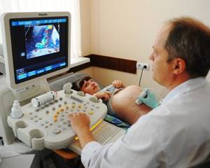
Ultrasound is a method for examining internal organs and tissues. It is carried out using an ultrasound machine equipped with a special sensor and monitor. The sensor is called a transducer.
When it comes into contact with the skin of a pregnant woman’s abdomen, it produces vibrations (sound waves), which it directs deep into the body.
There they collide with the woman’s internal organs (as well as with body parts and internal organs of the baby) and are reflected from them, subsequently displayed on the monitor.
Ultrasound can be two-dimensional, three-dimensional and four-dimensional. What's the difference between them? With a two-dimensional ultrasound, only vague outlines of the fetus are visible on the monitor in black and white. Thanks to a three-dimensional ultrasound, the doctor will be able to see a three-dimensional color image and examine the baby’s organs and systems in detail. Four-dimensional ultrasound allows you to see fetal movements.
This procedure is absolutely painless and does not create any discomfort.
Why is an ultrasound performed during pregnancy?
Carrying out research using ultrasound is not determined by the whims of future parents, but by the urgent need to diagnose various abnormalities in the child’s development and problems during the pregnancy itself.
Actually, the first ultrasound will be able to confirm pregnancy in the early stages, establish the presence of a fertilized egg (sometimes as many as two, or even three), and determine the exact duration of pregnancy. This method is very valuable in the presence of an ectopic pregnancy. After all, if you diagnose it early, you can avoid surgery.
Moreover, at a later date, ultrasound allows you to see whether pathologies exist. These include placental abruption, the threat of spontaneous abortion, and uterine hypertonicity.
Timely diagnosed pathologies and, accordingly, taken measures will allow you to maintain the pregnancy and give birth to a healthy baby. Ultrasound can also detect fetal malformations.
Sometimes they can be cured, and sometimes, unfortunately, the woman will be asked to terminate the pregnancy.
On the eve of childbirth, an ultrasound will allow you to find out some nuances that will determine both the course of the process itself and the behavior of doctors. Remember, only this method can tell with 100% accuracy whether there is an umbilical cord entanglement. And this is very important, since it threatens complications of the birth process, and sometimes creates a danger to the health, or even life, of the baby.
Do not forget that some women are required to undergo an ultrasound. And much more often than the majority of pregnant women. Such indications include chronic diseases. The most common among them are diabetes mellitus and various blood diseases.
You should not neglect the procedure if you have already had pregnancies that ended unsuccessfully (miscarriages, frozen pregnancies) or if there are seriously ill people in the family (for example, with Down syndrome).
When is the first ultrasound done during pregnancy?
A woman usually receives a referral for her first ultrasound during pregnancy at 12-13 weeks of pregnancy. This early research method is extremely important: with its help, the doctor will be able to assess the primary formation of the fetus and assess the process of the formation of organs and systems.
In some cases, the first ultrasound during pregnancy may be performed earlier. Firstly, to confirm the presence of pregnancy, and also to exclude the fact of an ectopic pregnancy.
Suspicion of an ectopic pregnancy requires mandatory examination using ultrasound - only in this way will it be possible to reliably establish whether this pathological condition actually occurs.
And only in this way will it be possible to intervene in time in the situation and carry out the necessary cleaning, otherwise serious consequences cannot be avoided.
The reason for an earlier ultrasound may also be alarming symptoms in the form of vaginal bleeding (or spotting) and nagging pain in the lower abdomen. Such symptoms most likely signal a threat of termination of pregnancy.
And, although it is very difficult to determine solely by ultrasound alone whether there is a threat of miscarriage, however, through such a study it is possible to find out the cause of the bleeding.
According to the ultrasound results, the doctor will be able to comprehensively assess the situation and give the woman appropriate recommendations.
Very often, a woman goes for an ultrasound, only suspecting pregnancy, without a referral from a gynecologist and of her own free will. Such actions are usually dictated by the desire to find out whether it is worth talking about pregnancy when characteristic symptoms are present, but the test does not show a result.
The question arises: at what stage will an ultrasound show pregnancy and is there any point in going for an examination at 1-2 weeks of delay in order to finally decide? The answer is yes: an ultrasound can show pregnancy already at 3-4 weeks, and this is precisely those 1-2 weeks of missed periods.
But it’s not always the case that if an ultrasound shows a fertilized sac in the very early stages, you can be guaranteed to be pregnant. Unfortunately, the fertilized egg may turn out to be empty and not contain an embryo, and this will only be possible to establish from the 5th week of pregnancy.
At what time are routine ultrasounds performed during pregnancy?
If the pregnancy proceeds favorably and without any abnormalities, the woman will be scheduled for three scheduled ultrasound sessions during the entire period of bearing the baby. The first ultrasound is performed in the first trimester, the second in the second, and the third, respectively, in the third trimester. A planned ultrasound during pregnancy allows you to assess whether everything is going “according to plan”, and, if there is suspicion, it is repeated.
The first planned ultrasound serves as a method for diagnosing pregnancy as such, allows you to determine whether there are risks of miscarriage, and diagnose “failures” in the development of the fetus at the earliest stages - when all vital organs and systems are formed and any deviation is fraught with the development of pathologies.
The second planned ultrasound during pregnancy is prescribed in the second trimester in order to assess the development of the baby, and at the same time the condition of the placenta. In addition, during the second ultrasound session, it is usually possible to determine the sex of the unborn baby.
The third planned ultrasound occurs, as you might guess, during the third trimester. At this stage, the degree of development of the baby, the state of the uteroplacental blood flow and even the presentation of the baby are traditionally examined.
The specialist who is managing the pregnancy will clearly answer the question of when to do an ultrasound during pregnancy. As a rule, the timing of ultrasound is determined as follows:
- first ultrasound – 10-14 weeks. The gestational age and approximate due date, the number of embryos are determined, and the tone of the uterus is assessed. The state of fetal formation, the likelihood of chromosomal abnormalities and developmental defects are also examined, the thickness of the cervical fold (neck area) is assessed - one of the main markers of Down syndrome;
- second ultrasound – 19-23 weeks. The sex of the baby, the size of the fetus and the correspondence of these indicators to the gestational age are determined. In addition to assessing the size and growth rate of the fetus, it is also possible to assess the development of the baby’s internal organs. In addition, the condition of the placenta, the amount of amniotic fluid is studied, and the absence of chromosomal abnormalities is confirmed;
- third ultrasound – 32-36 weeks. Necessary for diagnosing late fetal developmental anomalies that were not previously apparent. The size of the fetus is determined, and the date of the upcoming birth is specified once again. The condition of the fetus and its position before birth are assessed, and the possibility of entanglement with the umbilical cord is excluded.
Is ultrasound harmful to the fetus during pregnancy?
Most representatives of modern medicine unanimously assure that ultrasound is safe for the fetus and does not cause embryotoxic effects. Their opponents, representatives of different spheres of life, unanimously declare the allegedly monstrous consequences of using an ultrasound machine.
In fact, there is information that almost all “horror stories” are very exaggerated and do not have any serious evidence. Yes, ultrasound actually causes a slight heating of body cells, but this does not affect the condition and health of the fetus.
The connection between the use of ultrasound and various pathologies and anomalies of newborns has not been proven.
However, many question such arguments, saying that just because it has not been proven, this does not mean that it has no effect. Based on this, the following judgment will be reasonable: while scientists and doctors are researching this issue, we will be careful and once again we will not expose ourselves and the baby to ultrasound.
But if it is vitally necessary, that’s another question, because sometimes 10 minutes of examination save the baby’s life. Are any more arguments really needed? For those who are not convinced by this, we inform you: it has been proven that ultrasound for half an hour is safe for both the baby and the mother.
And the radiation, which opponents of ultrasound are so afraid of, actually lasts less than a minute. The rest of the time the device works for reception.
Especially for beremennost.net – Olga Pavlova
Source: https://beremennost.net/uzi-pri-beremennosti
How often can an ultrasound be done during pregnancy, is it harmful to the fetus?
Ultrasound diagnostics is a planned standard examination procedure during pregnancy. Ultrasound is considered one of the most reliable and safe procedures.
It allows you to diagnose fetal defects during intrauterine development, determine the condition of the child and the “uterus-placenta-fetus” system.
Thanks to ultrasound diagnostics, the obstetrician-gynecologist leading the pregnancy has the opportunity to intervene in the processes occurring in the mother’s body, and thus preserve the life and health of her and the child.
However, the question of the safety of this type of research continues to haunt the minds of future parents. How safe is the procedure for the baby? How often should an ultrasound be done during pregnancy? How many ultrasound procedures can be performed without health consequences? There is a lot of reliable information about ultrasound examination, but there is also a lot of speculation. It's time to figure out what is what.
Ultrasound diagnosis of pregnancy is by far the most informative study. It allows you to diagnose the development of the fetus, check all its systems and the readiness of the mother’s reproductive organs for delivery.
What is the ultrasound research method based on?
The method is based on the analysis of the difference between the signals of the probing and reflected ultrasonic waves. Using a special ultrasound sensor (transducer), an ultrasonic wave of 3.5 MHz is sent to the organ under study. Reflecting from various media and changing its frequency, the ultrasound wave returns and is absorbed by the receiver of the ultrasound sensor.
Next, the information goes to a computer, which is an integral part of the ultrasound diagnostic apparatus, which processes the received data. The ultrasound operator can see structures of different echogenicity on the monitor (amniotic fluid, fetal bones and tissues, etc.) and interpret the result.
The reliability of the information depends on the accuracy of the ultrasound machine and the experience of its operator.
Frequency of ultrasound procedures during pregnancy
How many times can an ultrasound diagnostic procedure be prescribed during the period of pregnancy? According to the order of the Ministry of Health of the Russian Federation No. 457 dated December 28, 2000, the algorithm for examining a pregnant woman includes 3 scheduled screening ultrasounds:
- the first screening in the period from 12 to 14 weeks is carried out to clarify the duration of pregnancy, its fertility and to check whether the size of the child and the thickness of the nuchal region correspond to the standards;
- the second screening in the period from 20 to 24 weeks is carried out to diagnose possible pathological processes in the “uterus - fetus - placenta” system, the place of placenta attachment, the condition of the child in the womb and its gender are determined;
- the third screening in the period from 32 to 34 weeks is carried out to clarify the presentation of the fetus, confirm the placenta insertion, as well as to confirm or remove suspicions of pathology and abnormalities of the child’s development. A comparative analysis of the child’s size and normative data is carried out, and the quantitative characteristics of the amniotic fluid are assessed.
The first screening is carried out at 12-14 weeks. It makes it possible to clarify the exact time of conception, compare the characteristics of embryo development with standards, and exclude genetic deviations
Reasons for prescribing additional ultrasound procedures
In addition to screenings, additional diagnostic ultrasound examinations may be prescribed, a referral for confirmation/clarification of previously identified pathology or by other methods, to monitor the condition of the mother and her baby and control the treatment process, if any, up to an ultrasound examination in the early stages. The reasons for ultrasound are quite compelling:
- burdened heredity;
- chronic diseases of the mother that can affect the development of the baby (diabetes, phenylketonuria, hypertension, etc.);
- when the expectant mother is exposed to pathological and harmful environmental factors (radiation, toxic substances, infections and intoxications);
- as a monitoring procedure for previously identified abnormalities during pregnancy.
Most expectant mothers wonder how long the ultrasound procedure takes, and how long will the fetus be exposed to ultrasound? The time required to complete the procedure depends on the type of study:
- standard ultrasound – 10 minutes;
- The duration of 3- and 4-dimensional ultrasound is from 30 to 50 minutes.
Ultrasound in the first weeks of pregnancy: benefits and harms
Reasons to prescribe an ultrasound early are:
There is no reliable data on the harmful effects of ultrasound in early pregnancy on the formation or development of the embryo, or any long-term consequences.
However, it is worth remembering that ultrasound is a medical diagnostic procedure. And it is carried out strictly according to indications. During an ultrasound, the fetus (its body and brain) is exposed to ultrasound radiation; this should not be done again.
How harmful is it to frequently conduct ultrasound diagnostics?
The harm of ultrasound during pregnancy has not been proven. No effects on child development were found. Of course, no doctor or scientist will give a 100% guarantee. Because it is difficult to conduct a pure experiment, i.e.
e. exclude the influence of other factors (examinations, treatment of the underlying disease, the influence of bad habits). It is also necessary to trace the effects of the ultrasound factor on several generations.
That is why they talk about relative safety or safety in comparison with other methods. Any medical procedure, any intervention should be carried out only if there is no other choice, especially in the early stages of pregnancy.
Of course, the embryo is exposed to radiation directed at it, and although the effect of such exposure has not been identified, it is better not to experiment.
In relation to any medical procedure, one must proceed from the following rule: when the benefit obtained from the procedure (in this case, ultrasound) exceeds the theoretically possible harm, then it should be carried out.
The baby in the womb reacts to external factors; during an ultrasound, he often shows activity and waves his arms. However, ultrasound has not been proven to cause harm or pain.
If you want to conduct an additional ultrasound, you should remember that not a single examination, including ultrasound, provides one hundred percent reliable information about the child’s condition. The reliability of the information is quite high, but it is still a probabilistic indicator.
Carrying out volumetric (3- and 4-dimensional) studies especially needs to be carried out strictly according to indications, because The time of exposure of the fetus to ultrasound waves during these procedures is significantly longer than with standard ultrasound.
The desire to photograph your unborn child or to clarify the sex of the baby is not a justification for an additional ultrasound procedure.
Myths about ultrasound diagnostics
Ultrasound examination is a breakthrough in non-invasive, painless diagnostic technology. How many ultrasounds are needed during pregnancy?
You should not refuse a fairly accurate and informative study, listening to the following unproven considerations:
- Ultrasound diagnostics negatively affects the genome.
Ultrasound allegedly deforms the DNA structure, causing mutations and developmental abnormalities in future generations. There is no data to support this theory. Experiments on mice have led to the denial of this hypothesis. - It is believed that the baby experiences pain when exposed to ultrasound, because
some children actively respond to exploration by waving their limbs. It is not known what the fetus reacts to: ultrasound, pressure from the transducer, the mother's excitement or her discomfort caused by a full bladder. If you pat your belly, your baby will most likely respond and push you back. This does not mean that your actions caused him suffering.
The examination must be ordered by a specialist, and the diagnosis must be carried out by a master of his craft using high-quality equipment. This minimizes possible risks and maximizes the undoubted benefits of ultrasound.
Answer to the question: “How often can an ultrasound be done during pregnancy?” - boils down to a simple recommendation: no more often than prescribed by the obstetrician-gynecologist leading the pregnancy.
Source: https://vedmed-expert.ru/prenatal/vredno-li-uzi-dlya-rebenka.html
How often can research be done?
The number of ultrasound procedures required to make a correct diagnosis is determined by the attending physician. To do this, you can do ultrasound frequently without worrying about your health. This has long been proven by modern science:
- sound waves do not have a negative effect on the body;
- ultrasound does not accumulate in tissues;
- Unlike X-ray examinations, ultrasound diagnostics has no contraindications and, if necessary, is performed on pregnant women and children.
As part of an annual preventive examination, women are recommended to undergo a mandatory ultrasound of the mammary glands and pelvic organs. In the presence of chronic and other diseases, the frequency of diagnostic examinations is determined by the doctor.
Experienced medical professionals can assure their patients that ultrasound can be done frequently if it helps timely detection of pathological changes in the body and speedy recovery. In turn, ultra-modern equipment allows us to guarantee high accuracy and reliability of the results.
At what time and how many times are ultrasounds performed during pregnancy?
Ultrasound during pregnancy is a safe examination of internal organs, which is prescribed to monitor the condition of the fetus and mother. During the entire pregnancy, a woman undergoes an ultrasound scan at least three times as part of screening - once in each trimester. There are indications for which the study is necessary outside of the schedule.
If certain pathologies of the fetus, uterus, cervix or placenta are suspected, ultrasound is prescribed on an emergency basis along with other diagnostic measures. Sometimes a fourth ultrasound is prescribed just before the birth. Most often it is carried out in the maternity hospital.
The timing of ultrasound during pregnancy is established for everyone, these are 10-12, 20-22 and 30-36 weeks.
Sometimes an ultrasound of the pelvic organs helps to understand whether a woman is pregnant. It is in the small pelvis that the uterus is located, where the chorion is fixed, as well as its appendages. Ultrasound can detect both normal and ectopic pregnancy.
This ultrasound is performed after 1-2 weeks of missed period. Pregnancy can be detected not only by the detection of a fertilized egg, but also by an increase in the thickness of the endometrium to 25 mm, as well as a large corpus luteum.
During a normal pregnancy, 3 scheduled ultrasounds are performed: at 10-12, 20-22 and 30-36 weeks. If a violation is suspected or to confirm the diagnosis, additional studies may be prescribed.
The most accurate results in this case are provided by transvaginal ultrasound. During the procedure, a special thin sensor is used, which is inserted into the vagina. A condom is first put on it. The sensor is very thin, pain and discomfort are excluded. It is located at a minimum distance from the uterus. It is completely safe and does not cause any threat of miscarriage or cause any discomfort.
The advantages of ultrasound in diagnosing pregnancy are the most accurate determination of timing. You can set the gestation time to the nearest day. This also allows you to accurately calculate your due date. Studies at 10-12, 20-22, 30-36 weeks do not have such accuracy due to the fact that each child develops individually in the womb.
Ultrasound in early pregnancy
At 10-12 weeks it is necessary to evaluate:
- The condition of the umbilical cord and make sure that there are two arteries in it;
- Cervix - measure the length;
- Place of attachment of the chorion in the uterus;
- Fruit size.
At this stage, the gestational age is clarified and possible pathologies are identified: Edwards syndrome, Down syndrome, Patau syndrome, abnormalities of the brain and other organs.
How is an ultrasound performed at 10-12 weeks?
The examination method is chosen by the treating gynecologist. Most often, the study is performed transabdominally - through the abdominal wall. The procedure algorithm is simple:
- The woman sits or lies down on the couch;
- A doctor or nurse applies a sound-conducting gel to the woman's abdomen;
- The doctor places the sensor on the abdomen and slowly moves it over the surface;
- The image is displayed on the monitor.
The procedure lasts a few minutes, after which the pregnant woman can immediately return to her usual activities. An ultrasound scan during pregnancy does not require any special preparation; it can be done at any time of the day.
For special indications, ultrasound is performed transvaginally. This is necessary when:
- Attachment of the chorion or placenta at a low level;
- The position of the fetus is difficult for measuring the collar area or other parts of the body;
- The need to assess the degree of isthmic-cervical insufficiency;
- Diagnosis of inflammation of the appendages or neoplasms in a pregnant woman.
Ultrasound at 21-22 weeks
The second ultrasound shows the size of the fetal body parts, its anatomy, and internal organs. Allows you to again clarify the duration of pregnancy, identify developmental delays, and find pathologies of the placenta, uterus, cervix, and umbilical cord. The maturity of the placenta is determined, premature aging is determined, and the amount of water is determined. At this time, the sex of the child is most often determined.
The second ultrasound is performed only transabdominally - transvaginal examination is not prescribed until after birth. The procedure is similar to the first ultrasound.
When is the last time an ultrasound is performed during pregnancy?
An ultrasound scan at the third screening is done at 30-31 weeks. As in the second study, the third ultrasound evaluates the condition of the uterus, placenta, umbilical cord, waters and the fetus itself.
In addition, it is necessary to determine the presentation of the embryo - the position inside the uterus. Normally it is cephalic, that is, the fetus lies head down, with the top of the head towards the exit.
It is important to establish the position of the placenta, the length of the cervical canal, and check the quality of the placenta.
The course of the third study does not differ from the course of the previous ones. An ultrasound is performed only on the abdominal wall.
Ultrasound before birth
Some women have an ultrasound scan upon admission to the maternity hospital. This is necessary to assess the condition of the woman and fetus and plan childbirth. The position of the embryo, height and weight, the condition of the cervix (maturity and degree of dilatation), the child’s heart rate, and blood flow in the vessels of the mother and fetus are taken into account. The study is also carried out only transabdominally.
Doplerometry during pregnancy
Doppler ultrasound during pregnancy is one of the types of ultrasound in which blood vessels are examined.
This type of ultrasound is prescribed to find out whether the baby has enough oxygen and nutrition. During a Doppler ultrasound, the blood flow in the vessels of the intrauterine fetus and the blood supply to the placenta and uterus are carefully studied.
Doppler is prescribed in the last trimester of pregnancy, regardless of the mother’s health condition. According to indications, Doppler may be prescribed earlier, at 20-24 weeks.
Reasons for prescribing additional Doppler ultrasound:
- Diseases such as diabetes, hypertension, gestosis, hypoxia.
- Unsatisfactory tests (often a cardiogram).
- Multiple pregnancy.
- Rhesus conflict.
- Pathologies of previous pregnancies.
- Inconsistency between fetal development and gestational age.